

UNIST Integrates Adaptive Optics Technology with Scanning-Enhanced Nanoscopy
 Published2021.06.17 11:35
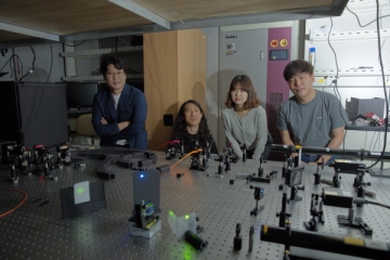
▲Professor Park Kyung-deok's research team (Professor Park Kyung-deok, top right)
An optical analysis technology has been developed that can freely change its observation method according to the subject, like a chameleon. It can selectively observe molecules bent in a specific direction or change the mode to detect optical signals from various substances. It is noteworthy that it can analyze the characteristics of ultrafine particles of various types, such as biological viruses, single chemical molecules, and semiconductor particles, all with a single microscope.
Professor Kyung-Deok Park's team from the Department of Physics at UNIST announced on the 17th that they have developed a new optical analysis technology that combines adaptive optics technology with a scanning-enhanced nanomicroscope.
The probe-enhanced nanomicroscope is a proprietary device that scans a sample with a sharp probe to obtain morphological information, while simultaneously analyzing the sample's optical properties by projecting the light collected by the probe onto the sample. Existing probe-enhanced nanomicroscopes were unable to control the polarization of the light transmitted from the probe to the sample, but adaptive optics technology was incorporated to overcome this problem. Adaptive optics adjusts the wavefront of light to compensate for wavefront distortion caused by scattering and other factors.
The gold probe of a probe-enhanced nanoscopy acts as an antenna, collecting an external laser beam. The light collected at the tip of the probe (the near field) is then focused on the sample, allowing various optical properties to be determined. This is because each sample has different light-responsive properties. The probe can also read morphological information, making it possible to simultaneously read the three-dimensional shape of a bent protein like a prions and changes in its chemical bonds.
On the other hand, existing probe-enhanced nanomicroscopes have a chronic problem: the polarization (direction) of light is fixed only perpendicular to the gold probe surface, making polarization control impossible. This makes it difficult to observe and select the alignment (orientation) of molecules. Even for identical molecules, chemical properties vary depending on whether they are lying or standing, making it crucial to be able to distinguish between them.
The research team solved this problem by incorporating adaptive optics, a method that uses computer algorithms to create customized laser beams. Unlike existing technologies that fix the wavefront of the laser beam directed at the probe, this technology adjusts the wavefront to match the probe's shape. This technology enabled free control of polarization direction with a resolution of 15 nanometers (10-9 m), and the technology was verified by distinguishing and measuring single molecules with different orientations.
Another advantage is the ability to obtain infrared absorption spectroscopic signals despite using visible-spectrum light from a laser beam. This phenomenon is possible because the gradient of the electromagnetic field changes rapidly over a very small space. Applying this, a single piece of equipment developed can selectively obtain visible-spectrum Raman and infrared absorption spectroscopic signals, depending on the intended use. A wide variety of microscopic particles can be studied using visible-light nanomicroscopy without the need for expensive infrared lasers and detectors.
Professor Park Kyung-deok said, “This study presents a new model of a fusion nano-microscope by combining adaptive optics, near-field optics, and computational imaging,” and added, “This study, which is the first to combine adaptive optics and near-field optics, which were studied independently, is expected to stimulate attempts to introduce adaptive optics into the field of near-field optics.”
Furthermore, adaptive optics technology was used to increase the efficiency of the laser beam focused on the probe, boosting the detection signal by over 200%. Because the intensity of light emitted from a space of approximately 10 nanometers is extremely low, enhancing the detection signal is crucial in nanomicroscopy.
Professor Park said, “Just as the development of telescopes has led to advancements in astrophysics, the development of new measuring equipment has led to the opening of new fields of research,” and expressed his will to conduct follow-up research with biomedical scientists, saying, “I would like to use the equipment we have developed this time for research on biomolecules such as coronaviruses and proteins.”
Meanwhile, Professor Jeong Moon-seok's team at Hanyang University participated in the production of the samples used in the study, and graduate students Gu Yeon-jeong and Kang Min-gu and undergraduate student Choi Jin-seong from the Department of Physics at UNIST jointly conducted the development and measurement research.
The research results were published in the international academic journal Nature Communications on June 8, and the original technology for the adaptive probe-enhanced nanomicroscope has been applied for domestic and European patents (PCT). This research was supported by the National Research Foundation of Korea and UNIST.
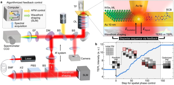
▲Schematic diagram and experimental schematic of the adaptive probe-enhanced nanomicroscope.

Comments
Related news
UNIST Develops Thin, Low-Power Quantum Dot TVs at Low Cost
A technique has been developed that applies high pressure using an ultrafine tip to control the brightness and wavelength of quantum dot light, and is drawing expectations for application in optical property analysis and display development.
2021.06.04by 배종인 기자

UNIST Dino Team Wins Hackathon
A zombie game created by UNIST students with their distinctive ideas and technical skills was selected as the best work at the hackathon.
2021.06.08by 강정규 기자

UNIST, Durability and Efficiency of Honeycomb-Type Thermoelectric Generators ↑
A research team at UNIST (President Lee Yong-hoon) comprising Professors Son Jae-sung and Chae Han-ki from the Department of Materials Science and Engineering, along with Professor Kwon Beom-jin from Arizona State University, has developed a technology to enhance thermoelectric generator durability and efficiency by 3D printing copper-selenide (Cu2Se), a thermoelectric material, into a honeycomb structure. The ability to produce complex honeycomb structures through 3D printing was made possible by newly developing an ink composed of thermoelectric material.
2021.06.16by 강정규 기자
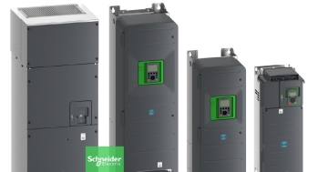
Schneider Aims for Building Energy Savings and Operational Stability with Next-Generation HVAC Drives
Schneider Electric has unveiled its next-generation HVAC drive product line 'Altivar HVAC'. The new product line's key features center on enhanced energy savings, system operational stability, and building management system integration. The products are divided into 'ATH200' for OEM and small equipment applications, and 'ATH600' for environments requiring continuous operation and advanced control. The company plans to expand the application scope from commercial buildings to facilities where HVAC system stability is critical, such as hospitals, airports, and data centers. The announcement includes over 30% energy savings, operation from -10°C to +60°C, Modbus and BACnet support, and compatibility with low global warming potential refrigerants.
2026.03.26by 명세환 기자
Ecolab Expands AI Data Center Cooling Business Through Acquisition of CoolIT
Ecolab has agreed to acquire CoolIT Systems, a liquid cooling company for AI data centers, for approximately $4.75 billion. The transaction is expected to close by Q3 2026, subject to regulatory approvals. CoolIT has been supplying direct-to-chip cooling-based coolant distribution units, cold plates, and manifolds, serving hyperscale data centers and colocation operators as customers. This acquisition is a measure to address the high-heat and high-density server environments created by AI proliferation. Ecolab plans to combine its water management and digital monitoring capabilities with CoolIT's hardware technology to expand its data center cooling business.
2026.03.26by 명세환 기자