


Ultra-fine 3D endoscope thinner than a hypodermic needle
 Published2022.09.02 10:47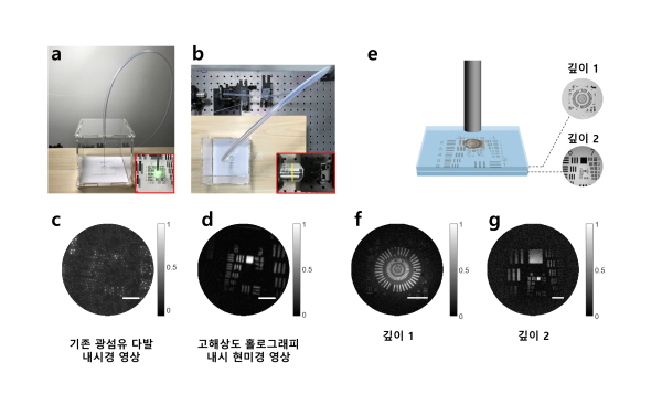
▲ Acquiring a 3D image of the inside of the box through a narrow tube
An ultra-fine endoscope has been developed that can observe capillaries and the nervous system in 3D.
A joint research team led by Deputy Director Won-Sik Choi (Professor of Physics at Korea University) of the Center for Molecular Spectroscopy and Dynamics (Director Min-Haeng Cho) at the Institute for Basic Science (IBS, President Do-Young No) and Associate Professor Young-Woon Choi of the Department of Biomedical Engineering at Korea University has developed an endoscope technology thinner than a hypodermic needle and succeeded in obtaining three-dimensional images of biological structures smaller than bacteria using this technology.
An endoscope is an imaging device designed to acquire images of objects in confined spaces or the inside of the human body.
Generally, endoscopes receive signals by inserting a thin imaging device into the object to be observed; they obtain images either by directly observing with a camera attached to the tip (probe) or by using optical fibers that transmit information using light.
When using a camera sensor, the probe becomes thicker, and in some cases, it may be necessary to make an incision in the skin to insert it. On the other hand, endoscopes using fiber optic bundles can be manufactured in a thinner form, which can minimize the incision site and patient discomfort.
On the other hand, conventional fiber optic endoscopes had difficulty obtaining clear images due to the empty spaces between the cores (the material that allows light to pass through the optical fiber) of individual optical fibers.
Furthermore, self-reflection at the ends of optical fiber bundles interfered with the observation of only the desired signal, making it difficult to observe biological structures with low reflectivity. Although fluorescent staining was required for observation, there were limitations to its application to the human body.
The research team overcame the limitations of existing fiber optic endoscopes by developing a very thin endoscope capable of high-resolution observation without attaching lenses or any equipment to the ends of the fiber optic bundles. The team focused light onto a single fiber within the bundle to illuminate an object located at a certain distance from the fiber.
Light reflected from an object transmits information about the object through multiple different optical fibers. A high-resolution image was obtained by measuring the resulting reflection holographic image and correcting for the distortion occurring in each core.
The developed endoscope has no equipment attached to the end of the optical fiber, resulting in a probe diameter of 350 μm (micrometers), which is even thinner than a hypodermic needle (approximately 500 μm). Using this, image information was obtained without fluorescent staining even from biological samples that are difficult to observe due to their very low reflectivity, such as mouse villi (structures within the small intestine).
In particular, the endoscope developed this time is capable of capturing microscope-grade high-resolution images, which is not possible with conventional fiber optic bundle endoscopes. It is capable of distinguishing objects even when they are separated by a distance of about 850 nm (nanometers). For reference, the size of bacteria is about 1,000 nm (1 μm, or one millionth of a meter). By correcting the measured holographic information, multi-depth 3D images can also be restored, and objects can be distinguished even when they are separated by a depth of about 14 μm.
Deputy Research Director Choi Won-sik stated, “We have developed a groundbreakingly thin, high-resolution endoscope,” adding, “This opens up the possibility of early disease diagnosis with minimal skin incisions, reaching areas that were difficult to access with conventional endoscopes, such as the lungs, capillaries, and even the brain and nervous system.”
The research results were published in the online edition of the international academic journal Nature Communications (IF 17.69) on August 2.

Comments
Related news
.jpg)
Kim Bit-nae-ri, IBS Director, selected as the first Korean member of the Royal Society of London
The director of the Center for RNA Research at the Institute for Basic Science (IBS, Director Noh Do-young), Kim Bit-naeri, Chair Professor at Seoul National University's School of Biological Sciences, has been selected as a Foreign Member of The Royal Society in the United Kingdom. This marks the first time a Korean has achieved this distinction.
2021.05.07by 배종인 기자
Sharing with the public the wondrous moments only scientists know about
기초과학연구원(IBS, 원장 노도영)은 제7회 IBS 아트인사이언스(Art in Science) 공모전을 개최하고, 6월7일부터 7월23일 17시까지 공모전 접수페이지(www.ibs공모전.com)를 통해 접수받는다. 개인(팀) 당 최대 3점까지 작품을 제출할 수 있다. 최종 수상작은 9월 발표 예정이다.
2021.06.07by 배종인 기자

IBS celebrates its 10th anniversary with a variety of events, including lectures by Nobel laureates.
The Institute for Basic Science (IBS, Director No Do-young) has organized various science concerts, including lectures by Nobel Prize laureates, to commemorate its 10th anniversary since its establishment.
2021.11.08by 배종인 기자

Korea Instrument, KOLAS Accredited Calibration Agency First to Simultaneously Acquire ISO/IEC 27,001·27,701
Korea Instrument has become the first among KOLAS accredited calibration agencies to simultaneously acquire ISO/IEC 27,001 and ISO/IEC 27,701 certifications. Through its proprietary platforms CWIZ Enterprise and CWIZ Partners, the company has embedded information security and personal data protection requirements across all business processes including customer information, equipment data, and calibration reports, with plans to continuously advance its management system through regular audits and security inspections.
2026.10.12by 배종인 기자

TeamViewer One, IT Incident Automation Workflow and Resolution Center Enhanced
TeamViewer has added IT incident automation capabilities and an advanced Resolution Center to its integrated digital workplace management platform 'TeamViewer ONE'. Recurrent service disruptions, configuration errors, and capacity shortages are automatically handled according to preset policies, and device status verification through script deployment can be managed in a unified interface, significantly reducing repetitive tasks for IT teams.
2026.10.12by 배종인 기자